Case Study: Life-Saving Bronchoscopy in a 2-Month-Old Infant with Severe Pneumonia
Department: Pediatric Pulmonology
Consultant: Dr. Siddhant Lalwani, Pediatric Pulmonologist
__________________________________________________________________________
Clinical Background:
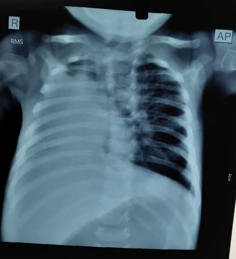
A 2-month-old infant was admitted to the hospital with severe pneumonia. The child had been under inpatient care for over two weeks with persistent symptoms and no significant clinical improvement. Chest X-rays showed progressive worsening of the right lung, and the infant remained dependent on oxygen support.
Clinical Presentation:
- Persistent fever and respiratory distress
- Oxygen dependency
- Radiological evidence of right lung consolidation
- No improvement despite aggressive medical management
Intervention:
Dr. Siddhant Lalwani, upon evaluating the infant, suspected an obstructive element and recommended a diagnostic bronchoscopy. Given the prolonged course, lack of response to treatment, and localized worsening on imaging, bronchoscopy was both a diagnostic and potentially therapeutic step.
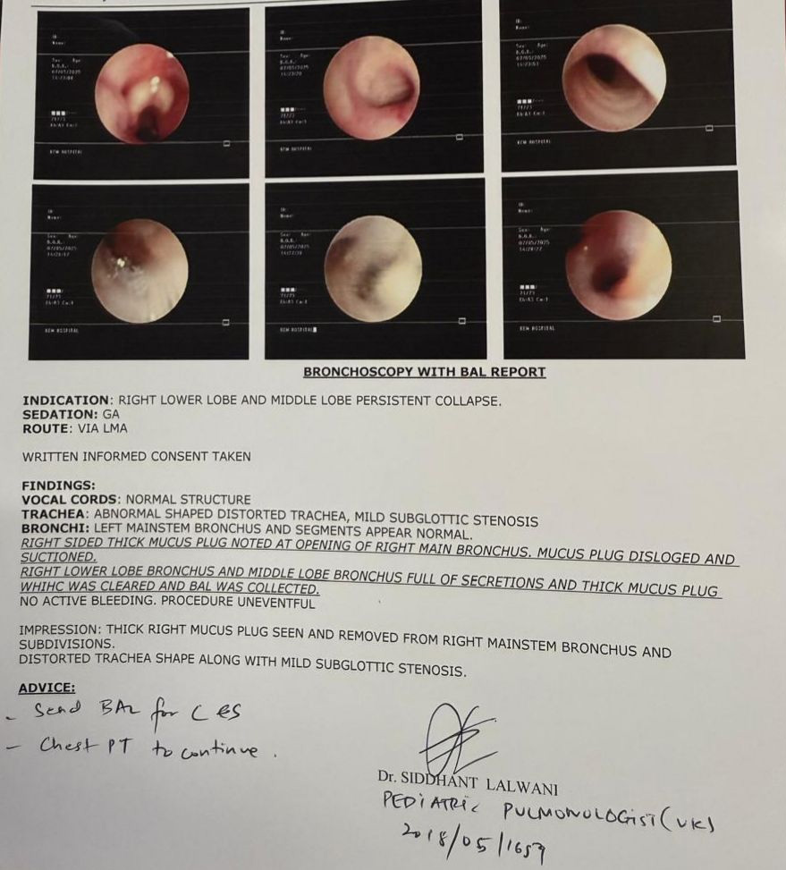
Procedure:
A flexible bronchoscopy was performed under controlled conditions. The procedure revealed a thick mucus plug completely obstructing a segment of the right lung. The plug was successfully extracted during the bronchoscopy.
Outcome:
- Immediate improvement in oxygenation
- Significant clinical improvement observed within 24-48 hours post-procedure
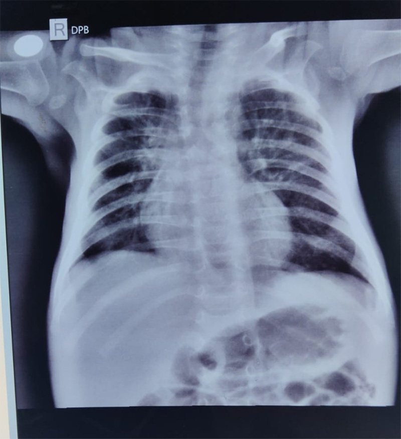
- Chest X-rays post-bronchoscopy showed clear re-expansion of the affected lung area
- Oxygen requirements were reduced
- The infant was gradually weaned off oxygen and is currently stable and recovering well
Conclusion:
This case highlights the critical role of bronchoscopy in both diagnosing and treating airway obstructions in infants. In this case, a simple yet timely intervention not only clarified the cause of persistent pneumonia but also provided immediate therapeutic relief.
Report